Spatial imaging, and virtual and augmented reality are impacting orthopedic surgery across all phases of care. By enabling detailed 3D exploration of patient-specific data and realistic surgical simulations, these tools improve professionals’ spatial understanding, precision, and decision-making.
Important notice regarding surgical planning and professional clinical use
Specialized functionalities for surgical planning and pre-operative professional applications are exclusive to Medical Imaging XR PRO FDA. This version is not yet available. Information about the release date will be published here soon.
Medicalholodeck is currently undergoing the required FDA (U.S. Food and Drug Administration) and CE (Conformité Européenne) certification processes. Our team is working diligently to ensure full compliance with all regulatory standards, and we expect Medical Imaging XR PRO to be available in both the United States and the European Union soon.
For updates on product releases, regulatory progress, and availability, or for any related inquiries, please contact info@medicalholodeck.com.
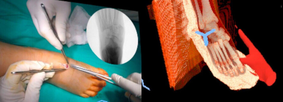
Presurgical planning in orthopedics
VR technology lets traditional 2D imaging transform into interactive, patient-specific 3D models. Surgeons can explore complex fracture patterns and anatomical relationships in an immersive environment supporting more precise surgical strategies and improving outcomes.
A recent study utilized a VR system integrating patient DICOM data with 3D trauma implant models that enabled surgeons to manipulate fracture and implant positioning in real time with accurate scale and spatial orientation. This hands-on interaction with both the patient's anatomy and implants within the same environment allowed detailed preoperative planning, potentially reducing operative time and fixation failure rates.
Read more
Minimal radiation exposure
Traditional imaging methods, such as Intraoperative radiography or fluoroscopy, generate ionizing radiation which poses potential health risks for patients and staff. Prolonged exposure can increase the likelihood of developing conditions such as cancer or cardiovascular disease.
VR and AR technologies offer promising solutions to mitigate these risks, since they can significantly reduce the number of intraoperative X-rays and fluoroscopy time. Simulated, risk-free environments allow surgeons and trainees to practice patient positioning and optimize the placement of imaging devices, such as the C-arm, which is crucial for acquiring high-quality images, while minimizing radiation exposure.
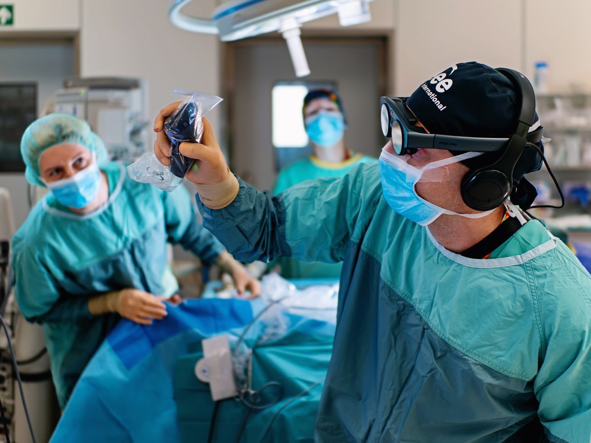
Intraoperative guidance
Real-time virtual reality collaborative session on the surgical treatment of Lisfranc injuries, enabling educators and trainees from around the world to participate. Medical Imaging XR (Medicalholodeck, Zurich, Switzerland).
Augmented reality serves as a navigation tool in orthopedic surgery. It has been shown to boost precision in placing screws, plates, or guidewires during fracture repairs, and to improve distal locking in intramedullary nailing. During shoulder arthroscopy, AR overlays enhance real-time visualization helping surgeons reduce errors and improve procedural efficiency. In total knee and hip arthroplasty, AR-assisted navigation reduces malpositioning risk, contributing to improved implant alignment, enhanced longevity, and lower complication rates.
Read more: https://doi.org/10.1002/ksa.12723
Integration of AI
The combination of artificial intelligence and virtual reality is transforming every step of orthopedic surgery, from preoperative planning to rehabilitation. AI algorithms analyze medical imaging to generate detailed, patient-specific anatomical models, while VR provides environments for surgical rehearsal and planning. This synergy improves diagnostic accuracy, optimizes implant selection, and allows virtual mock surgeries that train surgeons in a risk-free setting.
Intraoperatively, AI-driven systems can provide real-time feedback during procedures, optimizing implant alignment, and process live medical imaging to give suggestions minimizing surgical errors. While AI improves tissue recognition, VR technology further improves visualization through augmented reality overlays.
VR and AR combined with AI, allow surgeons to examine patient anatomy in detail, plan complex procedures, and minimize risks such as radiation exposure. They also support personalized rehabilitation through interactive therapy and continuous patient monitoring.
How to get started
Medicalholodeck integrates with secure hospital systems, providing PACS access, HIPAA-compliant data handling, and full patient data security. It runs on stereoscopic 3D displays, VR headsets, mobile devices, and standard Windows systems, enabling flexible use in hospitals, classrooms, and training centers.
Specialized features for surgical planning are exclusive to Medical Imaging XR PRO. Currently, Medicalholodeck is available only for educational use. The platform is undergoing FDA and CE certification, and we expect Medical Imaging XR PRO to be available soon in the U.S. and EU markets.
For updates, regulatory news, availability, or questions contact info@medicalholodeck.com.