Low-dose CT, high-impact:
VR is redefining pediatric surgery
Early detection and timely surgery are key for healthy lung development in babies. Low-dose CT scans reduce radiation but make images harder to interpret. Researchers at the University of Bonn created patient-specific 3D digital twins viewable in VR, improving visualization, spatial understanding, and collaborative decisions for the smallest patients.
The potential of Real-Time volume-rendered 3D Imaging in immersive virtual reality (VR) for surgical planning in infants with congenital thoracic malformation (CTM). Computational and Structural Biotechnology Journal.
Zalepugas D, Buermann J, Senkel S, Schmidt N, Ziegler AM, Kurz R, Schmidt J, Feodorovici P, Arensmeyer J (2025)
https://doi.org/10.1016/j.csbj.2025.11.005
Challenges of CT imaging
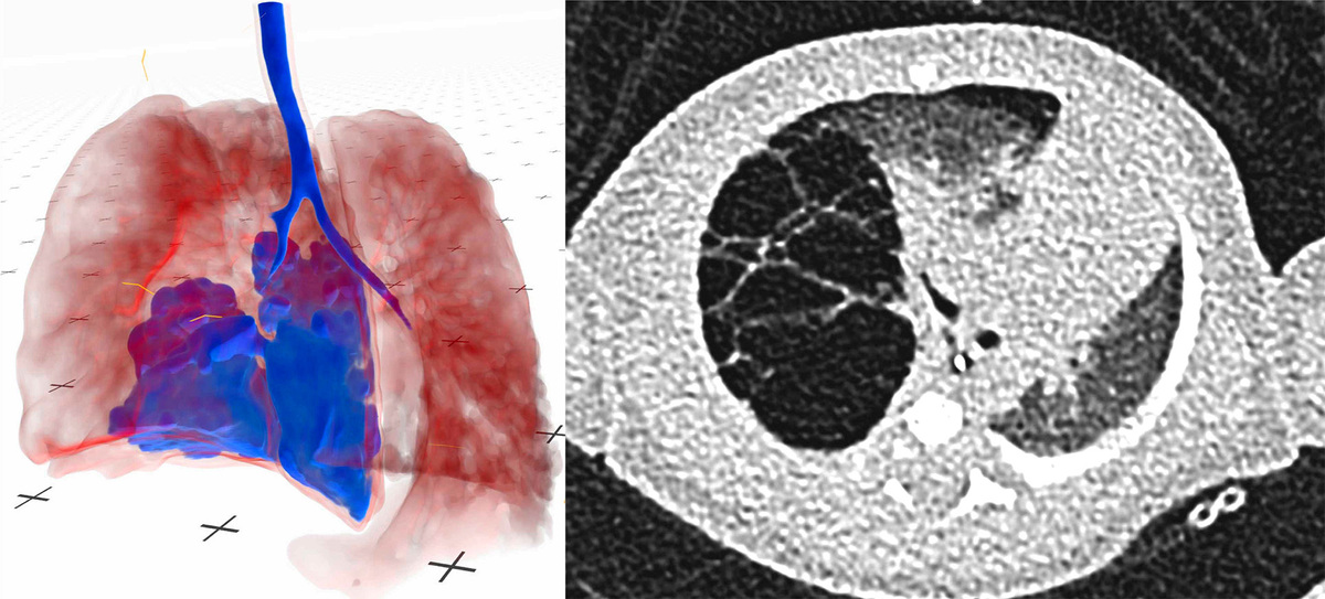
Congenital thoracic malformations (CTMs) include a wide range of rare lung anomalies that can be difficult to identify on low-dose CT scans, where subtle features and overlapping structures often obscure critical details. This makes accurate diagnosis and preoperative planning especially challenging, even though early surgical intervention is vital for healthy lung development in infants.
By leveraging VR-based 3D reconstructions, surgeons can gain a clearer, more intuitive understanding of each patient’s unique anatomy, improving spatial awareness and supporting better-informed surgical decisions.
Evaluating VR digital twins
This study aimed to determine whether live, volume-rendered 3D visualization of low-dose CT scans in virtual reality could enhance surgeons’ anatomical assessment and preoperative planning.
Four resident and four senior surgeons retrospectively evaluated 14 cases. Each case was first reviewed using conventional 2D CT scans with Xero Viewer (Agfa Healthcare, Mortsel, Belgium). After a three-month washout period, the same cases were reassessed using VR-based 3D models via Medical Imaging XR (Medicalholodeck AG, Zurich, Switzerland), allowing researchers to evaluate how VR influenced diagnoses and surgical planning.
Advantages of immersive 3D models
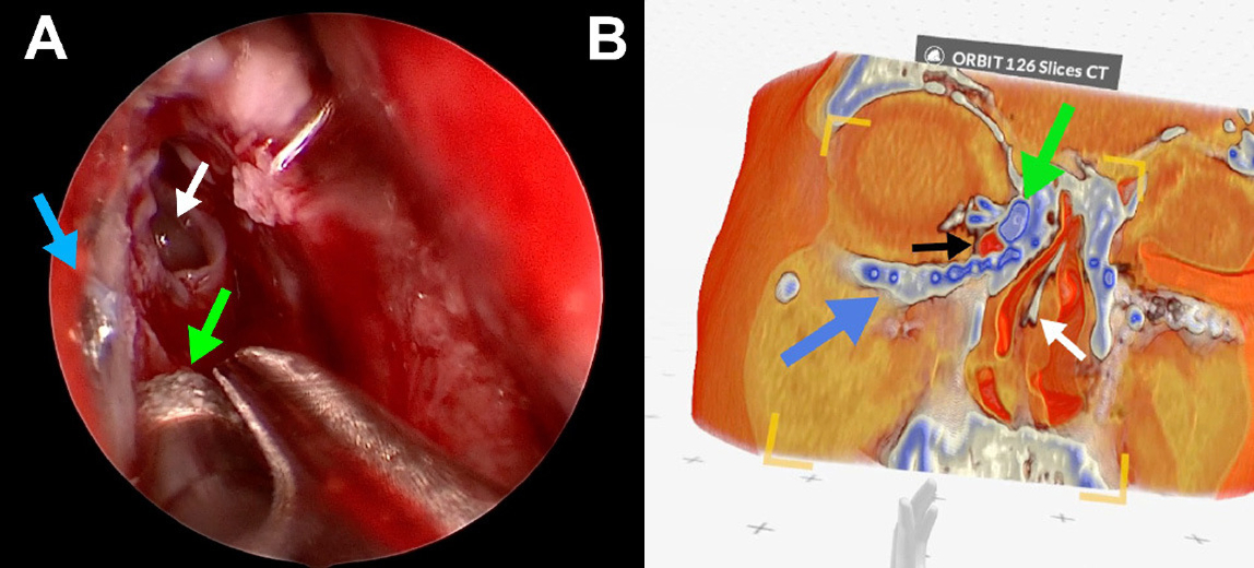
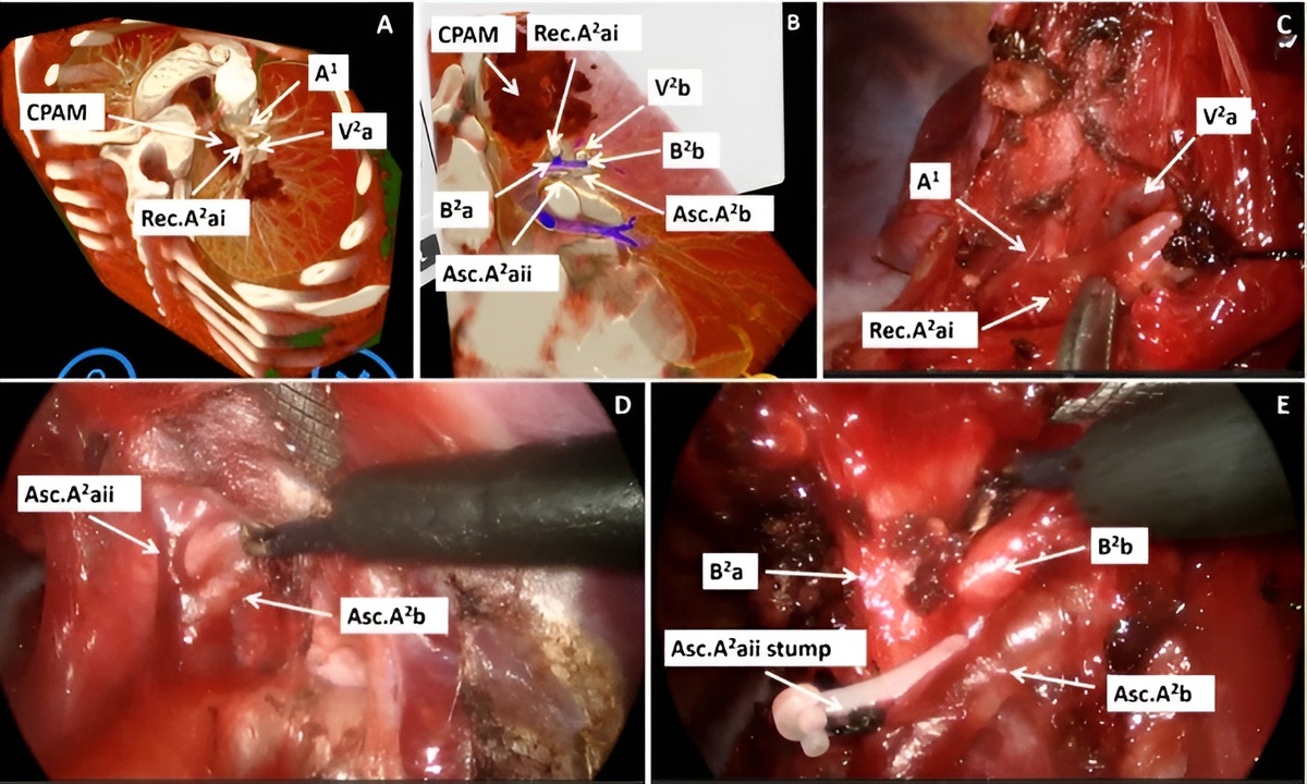
Immersive VR reconstructions of low-dose CT scans generate patient-specific digital twins, providing interactive 3D models with superior spatial insight compared to conventional 2D imaging.
While overall diagnostic performance was similar, VR revealed additional anatomical details not visible on standard CT. As a result, surgeons occasionally revised their diagnoses, with some changes later confirmed by pathology or intraoperative findings. In several cases, VR also uncovered a greater extent of disease than initially detected.
VR also influenced surgical planning, more frequently leading to lobectomy recommendations and supporting individualized, precision-medicine strategies.
Junior surgeons benefited particularly from VR, showing improved anatomical recognition and decision-making, suggesting it can help bridge experience gaps within surgical teams.
Expanding VR in surgery
Immersive VR-based 3D reconstructions extend beyond pediatric thoracic surgery. Patient-specific digital twins enhance spatial understanding, improving diagnostic accuracy and surgical planning in complex regions such as the heart, liver, and bones.
VR also benefits less experienced surgeons as a training and decision-support tool, helping standardize outcomes and reduce variability within teams.
Future steps include multicenter validation, integration into preoperative workflows, and functional digital twins combining anatomical and biophysical data to simulate patient-specific procedures. Haptic feedback can further enhance training, supporting precision medicine and effective team-based decision-making.
Check this out with Medicalholodeck
Medical Imaging XR enables clinicians to visualize, assess, and collaborate within an immersive, spatial environment. It allows real-time interaction with patient-specific 3D models, enhancing anatomical understanding, improving diagnostic accuracy, and supporting more precise surgical planning.
Medicalholodeck integrates with secure hospital systems, offering PACS access, HIPAA-compliant data handling, and full patient security. It works on VR headsets, PCs, iPads, and iPhones for flexible use in hospitals, classrooms, and training centers.
Specialized features for surgical planning are exclusive to Medical Imaging XR PRO FDA.
Currently, Medicalholodeck is available only for educational use. The platform is undergoing FDA and CE certification, and we expect Medical Imaging XR PRO FDA to be available soon in the U.S. and EU markets.
For updates, regulatory news, availability, or questions contact
info@medicalholodeck.com
November 2025